


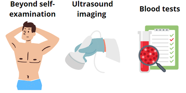
In recent years, Poland has witnessed a significant increase in the number of prostate and testicular cancer diagnoses. While this trend may seem alarming, it also highlights progress in preventive measures, diagnostics, and treatments. More men are being diagnosed at early stages, improving their chances for effective treatment and recovery.
Early detection is crucial for successful treatment and significantly increases the likelihood of a complete recovery.
Growing public awareness of the importance of regular screening is contributing to men being more willing to use medical services, resulting in better therapeutic outcomes.
However, if we have the misfortune and there is a need for in-depth diagnostics, medicine joins forces with chemistry and physics.
Imaging Technologies: A Journey Inside the Body
Imaging diagnostics are non-invasive methods that allow physicians to obtain detailed internal images of the human body. These techniques help detect organ or tissue abnormalities, including tumours, using radiation, sound waves, or magnetic fields.
Common Imaging Methods:
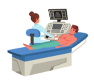 Ultrasound: This method uses sound waves to create images of internal organs. Ultrasound is often employed to diagnose breast, thyroid, and abdominal cancers and in prenatal imaging.
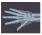 X-rays: An older, widely recognized imaging technique useful for diagnosing cancers, fractures, and other conditions. X-rays are essential for assessing bone structure and some internal organs.
 Computed Tomography (CT): A CT scan involves taking a series of X-rays from different angles, which are then combined into a 3D image. CT scans are pivotal in determining tumor size, location, and disease progression.
 Magnetic Resonance Imaging (MRI): This technique uses powerful magnetic fields and radio waves to produce highly detailed images of soft tissues, making it indispensable for diagnosing brain, spinal cord, and some internal organ cancers.

PET, or positron emission tomography: this is an advanced medical imaging method that allows doctors to ‘see’ what is going on inside our bodies at a cellular level, and more specifically allows the metabolic activity of cancer cells to be assessed. A radioactive tracer is administered to the patient, which accumulates in the cancer cells. This allows the size of the tumor, its metabolic activity, and the spread of the disease to be assessed.
Now that we know so simply, without going into complicated mechanisms, how the tests in question work and what they consist of, let us consider in more detail where this involvement of chemistry in diagnostic tests is.
In the description of PET, the tricky phrase ‘radioactive tracer’ appears, but there are more such helpers, which are able to exotify the studied image or, like a highlighter, indicate a specific disease area. And these little helpers are so-called cotrast agents.
Contrast Agents: Enhancing Diagnostic Precision
Imagine that you want to see what is at the bottom of an opaque glass. You can add a special dye to it - then what was previously invisible will become clearer. A contrast agent in imaging studies works similarly. This is a special substance that, when introduced into the body, allows doctors to obtain more detailed and clearer images of internal organs during imaging examinations. This makes it easier to diagnose various diseases, including cancer.
Contrast agents are specialized substances introduced into the body to improve imaging clarity. They act like highlighters, pinpointing disease-affected areas during diagnostic scans.
Applications of Contrast Agents:
- CT scans: To evaluate internal organs, circulatory systems, and urinary systems.
- MRI: For imaging the brain, spine, joints, and muscles.
- X-rays: For assessing the digestive and urinary systems.
Depending on the specific imaging method, different contrast agents are used. In X-ray-based techniques, contrast agents typically contain iodine or, less commonly, barium sulfate. These agents absorb external X-ray radiation, reducing its intensity on the detector. Simply put, contrast agents absorb radiation, resulting in darker areas on the generated images. This mechanism differs from radiopharmaceuticals used in nuclear medicine, such as in PET scans, where compounds containing radioactive elements emit radiation.


In MRI, contrast agents, most commonly gadolinium-based, work differently. They alter the magnetic properties of tissues they infiltrate, leading to variations in signal intensity on the images. This distinction enables better visualization of pathological changes, such as tumors or sclerosis.
How Does It Work?
Imagine the human body as a collection of tiny magnets. Inside the MRI scanner is a powerful magnet that aligns these tiny magnets (protons in hydrogen atoms) like compass needles. The scanner emits energy impulses that make these magnets spin and wobble. Each tissue reacts differently to these impulses, producing the distinct shades and colors visible in MRI images.
Contrast agents in MRI act like a magnetic storm, disrupting the alignment of these compass needles. This disruption is detected by the scanner and converted into an image. Healthy tissues, with minimal disruption, appear darker, while areas with higher contrast agent accumulation, such as tumors, show brighter regions.
Why Gadolinium?
 Gadolinium possesses unique magnetic properties that interact with the strong magnetic field of an MRI scanner, modifying signals from the tissues it enters. This enables the detection of pathological areas with higher precision. Gadolinium-based contrast agents are typically administered intravenously or orally, depending on the imaging target, such as the gastrointestinal system.
To ensure safety, gadolinium is chemically bound to organic molecules, forming stable complexes. These compounds resemble nanostructures with a protective coating, determining their properties and suitability for specific imaging targets, such as liver, cartilage, or brain tissues.
Criteria for an Ideal Contrast Agent
An ideal contrast agent must meet stringent safety and performance standards:
- Low Toxicity: It should be minimally harmful to the human body.
- Efficient Elimination: Rapidly excreted, primarily through the kidneys, reducing adverse effects.
- Minimal Allergic Reactions: A lower likelihood of hypersensitivity increases its safety profile.
The ideal contrast agent should be safe, effective, and easy to use. Thanks to continuous research and technological developments, contrast agents used in imaging studies are becoming increasingly safe and effective. Such compounds are as thoroughly tested as drugs, so they must meet several restrictions.
And are new compounds being worked on?
Despite the widespread use of gadolinium-based agents, they have limitations, such as the potential retention of gadolinium in some patients. This has spurred research into alternatives, including iron-based compounds.
Key Challenges:
- Safety: Any new contrast agent must ensure patient safety without severe side effects.
- Efficacy: It must provide high-quality imaging to facilitate accurate diagnoses.
- Stability: The agent should remain chemically stable and resist breakdown in the body.
- Cost: Developing and manufacturing new agents is an expensive and time-intensive process.
Cutting-Edge Research: Iron-Based Contrast Agents
At the forefront of innovation is the team led by Professor Nikodem Kuźnik, the only group in Europe dedicated to synthesizing modern iron-based contrast agents. Their groundbreaking work is globally unique, rivaled only by two research groups in the United States.
Final Thoughts
Prioritizing regular health check-ups reduces the need for advanced diagnostic tests. When such tests become necessary, contrast agents enhance their accuracy, guiding precise diagnoses. With ongoing chemical advancements, the future of contrast agents looks promising, paving the way for safer and more effective imaging solutions.